The digestive system of the cat is made up of a collective network of organs designed to supply the body with the nutrition it needs for growth, maintenance, and repair.
It also functions to rid the body of waste. Because of this, diseases involving the digestive system can have a profound effect not only on the region so afflicted but on the entire body as well.
Anatomy and Physiology
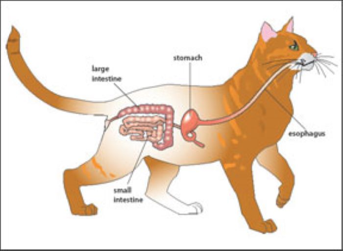
Those organs or regions of the body categorized under the digestive system include the oral cavity (teeth, tongue, salivary glands, etc.), esophagus, stomach, small intestine, large intestine, rectum, anus, pancreas, liver, and gallbladder. In order to understand diseases and their adverse effects, a brief overview of the digestive process is warranted.
Keep in mind, however, that the following description is an oversimplification; the actual digestive process is so complex and involved that it warrants the devotion of an entire book in itself! Once food enters the oral cavity, the process of digestion begins.
The teeth mechanically rip, crush, and grind down the food, while the saliva secreted by the salivary glands into the mouth moistens and partially digests carbohydrate portions of the food before it is swallowed.
In kittens, the eruption patterns of these teeth do not start until around 2 to 4 weeks of age. By the time the neonate is 7 weeks of age, it should have a full complement of deciduous (baby) teeth. By 7 months of age, all the permanent teeth should have fully erupted and replaced the deciduous ones.
From the oral cavity, food, water, and saliva are passed back into the esophagus with the aid of the tongue and are swallowed. The walls of the esophagus consist of bands of muscle, which contract in a rhythmic fashion, pushing the food down toward the stomach.
This unique muscular action is called peristalsis. From the esophagus, the food passes through a muscular sphincter into the stomach. This sphincter is very important, for it keeps stomach acids and enzymes from entering into and burning the esophageal lining. If it is defective, ulcers and that feeling humans describe as “heartburn” can result.
The stomach is lined with cells that secrete acids and special digestive enzymes, designed to further break down ingested proteins and carbohydrates. The muscular walls of the stomach help gently churn and mix the contents, until such time as they’re ready for passage into the small intestine.
Again, the food must pass through another sphincter to reach the small intestine. Once the food passes through the sphincter, the stomach acids mixed in the digesta are neutralized and rendered harmless by secretions in the small intestine.
The small intestine is the site where most digestion occurs, and where the resulting nutritional building blocks are absorbed into the body. Bile produced by the liver and stored in the gallbladder is added to the digesta here to break down fats.
Enzymes from the pancreas further digest fats, proteins, and carbohydrates until, finally, the nutrients can be absorbed through the intestinal lining and into the body. The lining of the small intestine consists of millions of small, fingerlike projections called villi, designed to increase the absorptive surface area within the intestine.
And as if this weren’t enough, each of these tiny villi are lined by even tinier projections, called microvilli— again, to further increase the surface area for nutrient absorption. One reason why a parvovirus infection can be so deadly is that the virus can effectively destroy these villi lining the small intestine, blocking absorption of nutrients and “starving” the victim.
As digested nutrients are absorbed, they enter into either the circulatory system or into the lymphatic system. End products of protein and carbohydrate digestion travel by way of the blood to the liver, where any toxic by-products are promptly eliminated.
Other functions of the liver include the production of serum proteins, the storage of vitamins and other nutrients, the destruction and removal of old red blood cells from the bloodstream, and the secretion and excretion of bile into the small intestine to aid in the digestive process.
Fatty acids, resulting from fat digestion, travel initially via the lymphatic vessels and later enter into the bloodstream, where they are broken down and absorbed into the body tissues. The lymphatic system also plays an important role in immunity.
Digesta and waste that are not absorbed from within the small intestine then pass into the large intestine, which is responsible for removing water and electrolytes from the material and lubricating it for passage out of the body through the rectum and anus.
One special portion of the large intestine is called the cecum, which corresponds to the human appendix.
Debbie Reid is the founder and chief editor of CatPedia.net, with over 25 years of hands-on experience in feline care, rescue, and behavior research. Inspired by her rescue cats Snickers and Bigboy, she collaborates with animal welfare resources and consults veterinary literature to turn complex feline health, nutrition, and behavior topics into clear, actionable guides.