Otitis media, infection involving the middle ear, usually results from a chronic, untreated or recurring otitis externa. In such cases, the eardrum might become so diseased as to tear or rupture completely, allowing direct access of infectious organisms into the middle-ear chamber.
The clinical signs of otitis media are essentially the same as those for otitis externa, with a few notable additions. Pets so afflicted will usually exhibit a head tilt toward the side of the affected ear. In severe cases, paralysis of the facial muscles on the side of the lesion might be seen as the nerves passing through the middle ear become involved.
This can result in a characteristic drooping of the eyelids, cheeks, and lips. In addition, a decreased tear production, pinpoint pupil, and protrusion of the third eyelid might be noted in the eye on the affected side.
If the infection extends from the middle ear into the inner-ear apparatus, the signs become even more pronounced.
Since the inner ear functions in maintaining balance and equilibrium as well as hearing, pets suffering from otitis interna tend to become very uncoordinated and might fall down frequently or move in circles toward the affected side.
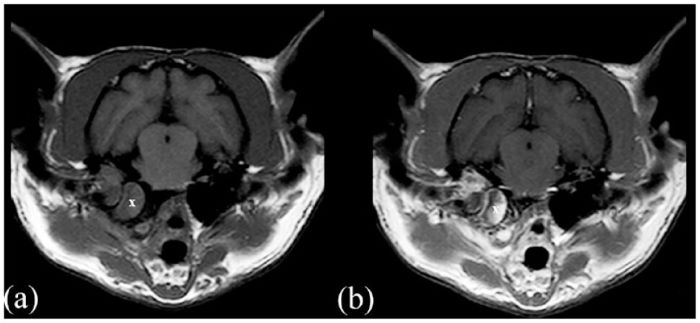
A characteristic twitching of the eyeball, called nystagmus, also becomes more noticeable. Although the clinical signs seen are often diagnostic, radiographs of the skull are quite helpful at confirming a diagnosis of otitis media or interna and determining the extent of the disorder.
Therapy for otitis media or interna must be instituted promptly to prevent permanent damage to the hearing apparatus. Oral antibiotics should be started immediately. In cases of otitis interna, continued treatment with antibiotics might be required for up to 30 days to afford a complete cure.
In select cases, anti-inflammatory medications have been used to reduce signs associated with inflammation. If not already ruptured, the eardrum on the affected side is usually punctured to allow for thorough drainage of the middle-ear cavity and for the direct infusion of medications.
Of course, such treatment steps must be carried out in a veterinary hospital under heavy sedation or anesthesia. In tough, refractory cases, surgical placement of a drain in the bony tympanic bulla affords excellent exposure to the middle- and inner-ear spaces.